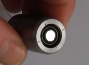
Retinoscopes are a very common optical instrument used by optometrists to examine a patient’s eye and evaluate the need of corrective lenses. They use a very interesting physical principle.
Let’s first describe the optical instrument. The retinoscope is a hand held device that the optometrist placed right in front of their eyes. The retinoscope projects a slit into the patient’s eyes and the optometrist will look at the red reflex that illuminates the pupil to determine the required accommodation for the patient. The red reflex is what we usually think of as “red eye” when taking a picture with flash.

Figure 1 shows the use of a Retinoscope from the patient’s point of view (A) and a lateral view (B). Image from Survey of Ophthalmology
By rotating the retinoscope, the slits are moved across the pupil. IF the patient’s far point lies behind the observer’s eye the observer will see that the red reflex moves in the same direction as the motion of the slit. This movement is called “with motion”. Likewise, if the patient’s far point lies between the observer and the patient the red reflex with move in opposite direction to the slit’s movement (or “against motion”). Finally, when the patient’s retina is conjugate to the retinoscope we will have a motionless uniform illumination (also called “point of neutrality”).

Figure 2. Red reflex movement with respect of the slit movement. Image from Westmeadeye.com
The optometrist can place lenses with different powers in front of the patient’s eye until the optometrist find the point of neutrality.
Optical design
From the point of view of optical design. A basic retinoscope can have a very simple configuration. It will have a light source, lens, and a movable barrel that can change the distance between the source and the lens. Additional elements include cards with the slit profile and focusing cards that helps the patient direct their sight into the device.
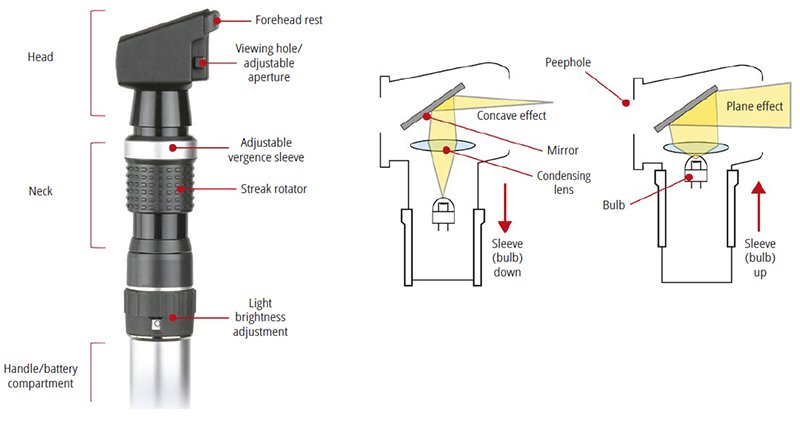
Figure 3. Basic configuration for a Retinoscope
Retinoscopes are very useful when working with small children. Because it’s a less subjective test and less intimidating that using a traditional Phoropter.
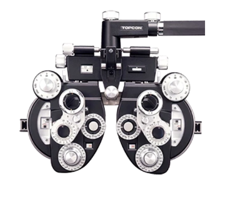
Figure 4 Traditional Phoropter can be intimidating for small children.
